|
Home Anatomy | Arthritis | Partial Knee Replacement | Total Knee Replacement | What to Expect on Surgery Day | Post Operative Care | Knee Rehabilitation | Non-Surgical Alternatives | Life After Knee Replacement |
Knee AnatomyThe following article will provide knee anatomy information for patients and their families.The largest joint in the body, the knee is also one of the most complex. The knee may be described as a modified hinge joint, similar to the hinge on a door. However, the knee not only bends back and forth like a hinge, it has a complex rotational component that occurs with flexion and extension of the knee. The knee is a major weight-bearing joint that is held together by muscles, ligaments, and other important soft tissue. Cartilage is the material inside the joint that provides shock absorption to the knee during weight-bearing activities such as walking or stair climbing. Below is an illustration of knee anatomy with its major bones, ligaments and muscles appropriately labeled. 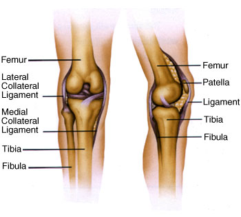 The bones of the knee are the femur (thigh bone), tibia (shin bone) and patella (kneecap). The femur and tibia meet to form a hinge with the patella in front of these two bones protecting the joint. The patella slides up and down in a groove in the femur (the femoral groove) as the knee is bent and straightened.
Knee Anatomy: Ligaments
Knee Anatomy: Cartilage
Knee Anatomy: Tendons Bones, ligaments, cartilage and tendons all work together to build a healthy knee. Click on the image below to view an interactive animation of knee anatomy.(136k - Requires: Requires Free Flash Player, Download Here) 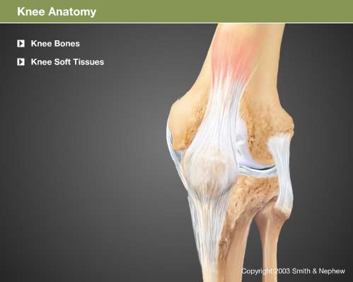 |
|
Contents Courtesy of www.knee-replacement-info.com |